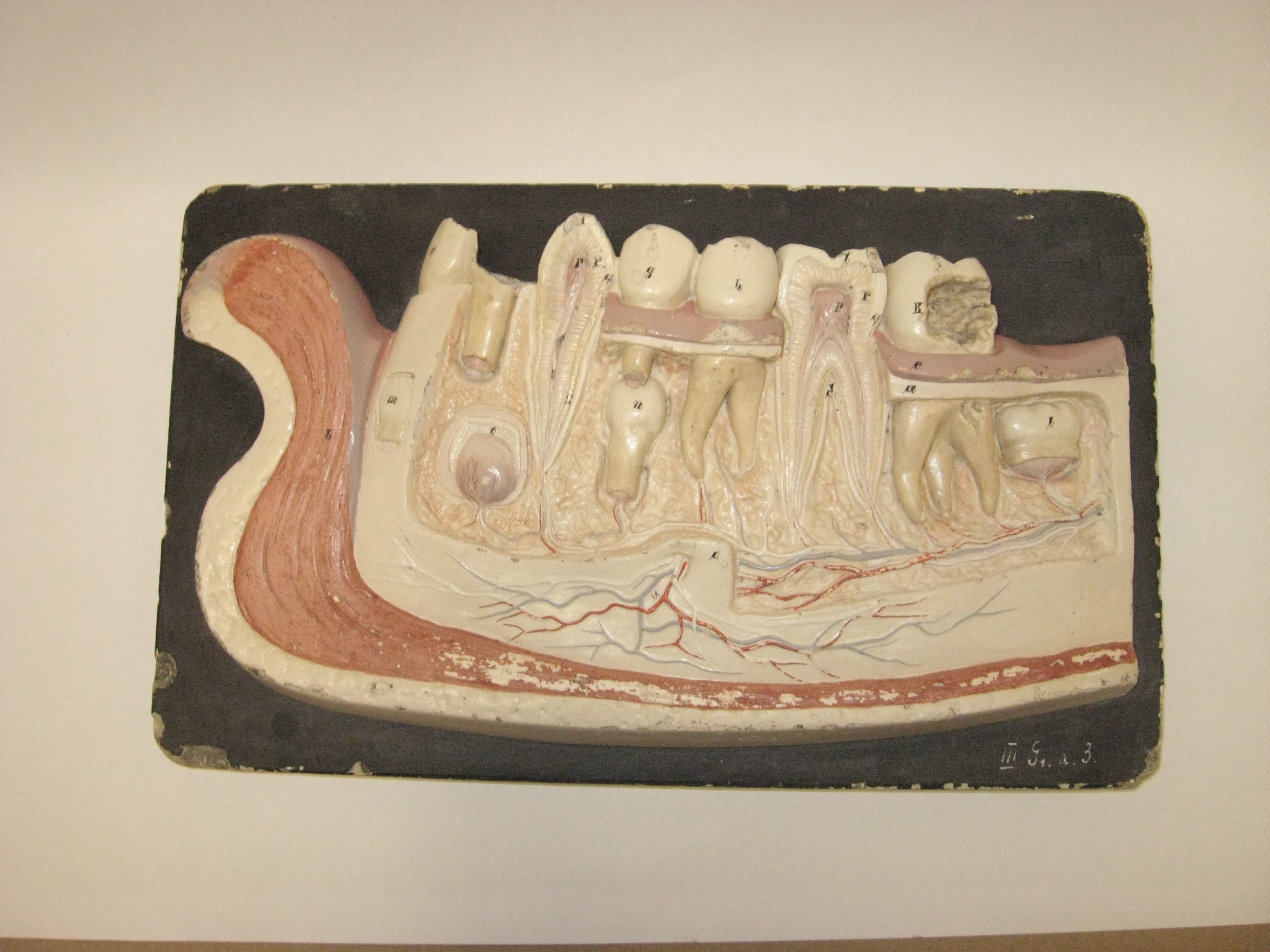
Odontological model, anatomical dental model, teaching aid for anatomy. Dental model or dental model. Anatomical model of the mandible. This teaching aid comes from the natural science laboratory of the Second State Gymnasium in Przemyśl, Zasanie. The model was brought by Professor of Natural Science and Natural History, Julian Kubrakiewicz, who ran the natural science laboratory from 1921 to 1931, at the Second State Gymnasium (from 1922, the Second Gymnasium named after Professor Kazimierz Morawski in Przemyśl). This and other models were used for anatomy instruction. A plaster model of the human mandible, showing the anatomy and position of the teeth in the lower right arch, is a rectangular, varnished plaster plaque. The specimen shows a cross-section of the lip, the bone layer, and the types of teeth, their external and internal structures. The incisors (one broken off), molars, and premolars, with cross-sections, are cream-colored. The gums are pink. The blood vessel threads are red, and the nerves are gray. Tooth buds, the method of tooth formation, and tooth pathology are also shown. The process of replacing primary teeth with permanent teeth is illustrated illustratively. Several anatomical structures are numbered. The model was created in a German workshop at the University of Leipzig, where sculptor Franz Josef Steger (1845-1938) collaborated with pathological anatomist Carl Ernst Bock (1809-1874).
Odontological plaster model