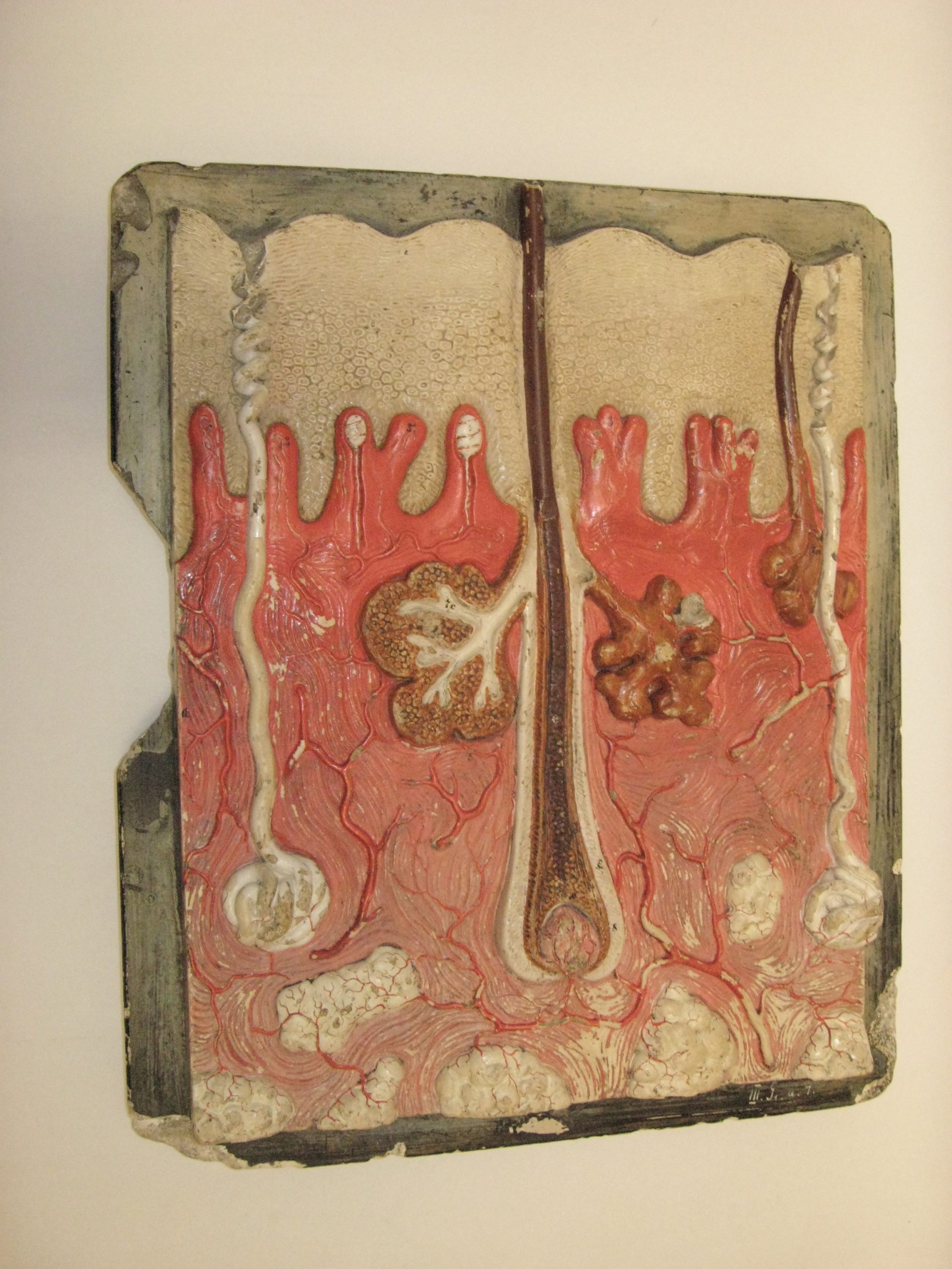
Anatomical model of human skin. A block model of a sagittal section of a fragment of human scalp, presented on a rectangular plaster plaque (a chip is visible on the left edge). The base is black, with the relief painted in red, brown, and cream. This teaching aid comes from the natural sciences laboratory of the Second State Gymnasium in Przemyśl, Zasanie. The model was brought by Professor of Natural Science and Natural History, Julian Kubrakiewicz, who ran the natural sciences laboratory from 1921 to 1931 at the Second State Gymnasium (from 1922, the Second Gymnasium named after Professor Kazimierz Morawski in Przemyśl). This and other models were used for teaching anatomy. The model reproduces the appearance of the human scalp at 80x magnification, with an actual thickness ranging from 3.5 to 5.5 mm. The scalp is one of the thickest parts of the human body. The cross-section shows all layers. The epidermis, dermis, and subcutaneous tissue are visualized in two-dimensional detail. The structure of the hair, including the root, bulb, papilla, follicle, and shaft, is highlighted in brown. Above the hair follicle, the sebaceous glands are visible (one in the cross-section). The sweat glands are marked in creamy white. The blood vessel strands are colored red. Several anatomical structures are numbered. The model was created in the German studio at the University of Leipzig by sculptor Franz Josef Steger (1845-1938), who collaborated with anatomist-pathologist Carl Ernst Bock (1809-1874).
Plaster Skin Model